
Two-month-old babies are already making sense of the world
New research from neuroscientists at Trinity College Dublin shows that babies as young as two months old can already
New research from neuroscientists at Trinity College Dublin shows that babies as young as two months old can already organize what they see into distinct object categories. This ability appears far earlier than scientists previously believed and suggests that important building blocks of perception are present almost from the very start of life.
By combining brain imaging with artificial intelligence models, the researchers gained new insight into how infants think and learn during their earliest months. The findings help clarify what is happening inside a baby’s brain long before speech or deliberate movement is possible.
The study was recently published in the journal Nature Neuroscience by a research team from the Trinity College Institute of Neuroscience (TCIN) and the School of Psychology.
What Scientists Are Learning About the Baby Mind
“Parents and scientists have long wondered what goes on in a baby’s mind and what they actually see when they view the world around them. This research highlights the richness of brain function in the first year of life,” explains Dr. Cliona O’Doherty, the study’s lead author, who carried out the work while based in Trinity’s Cusack Lab.
“Although at two months, infants’ communication is limited by a lack of language and fine motor control, their minds were already not only representing to how things look, but figuring out to which category they belonged. This shows that the foundations of visual cognition are already in place from very early on and much earlier than expected.”
Studying Awake Infants With Brain Scans
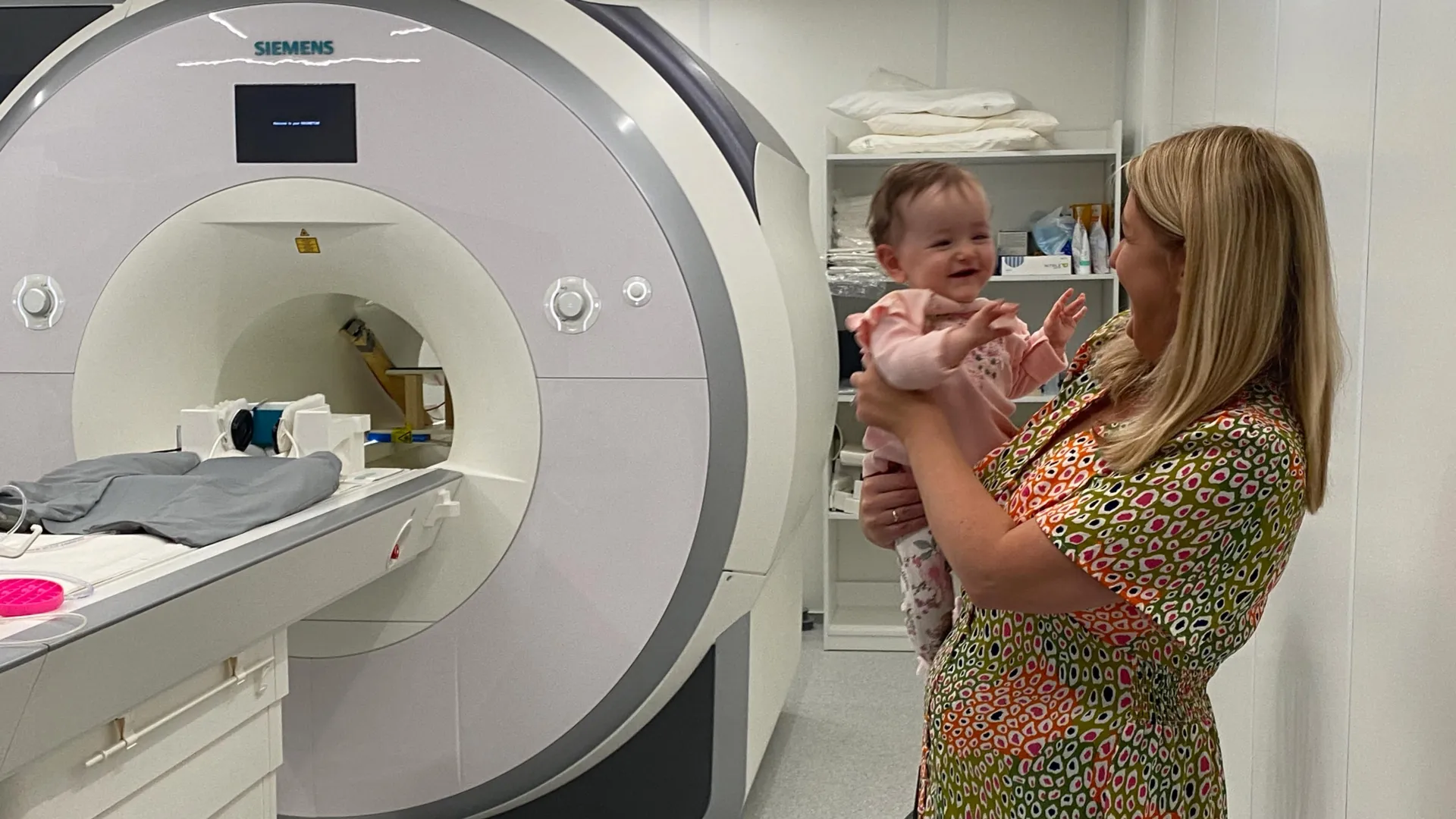
Working with the Coombe and Rotunda Hospitals in Dublin, the FOUNDCOG team recruited 130 infants who were all two months old. Each baby lay comfortably on a soft beanbag while wearing sound-cancelling headphones and viewing bright, colorful images designed to hold their attention for 15-20 minutes.
This setup allowed researchers to use functional MRI (fMRI) to record patterns of brain activity as the babies looked at images from 12 familiar visual categories such as cat, bird, rubber duck, shopping cart and tree.
How Artificial Intelligence Helped Decode Brain Activity
After collecting the brain scans, the team used artificial intelligence models to analyze how different visual categories were represented in the infant brain. By comparing activity patterns along visual recognition pathways in the models and in the babies’ brains, the researchers could better understand how early categorization takes place.
“This study represents the largest longitudinal study with functional magnetic resonance imaging (fMRI) of awake infants. The rich dataset capturing brain activity opens up a whole new way to measure what babies are thinking at a very early age. It also highlights the potential for neuroimaging and computational models to be used as a diagnostic tool in very young infants,” says team leader Rhodri Cusack, the Thomas Mitchell Professor of Cognitive Neuroscience at Trinity’s School of Psychology and Trinity College Institute of Neuroscience.
“Babies learn much more quickly than today’s AI models and by studying how they do this, we hope to inspire a new generation of AI models that learn more efficiently, so reducing their economic and environmental costs.”
Why These Findings Matter Beyond the Lab
Dr. Anna Truzzi, now based at Queen’s University Belfast and a co-author on the paper, emphasized how recent advances made the research possible. “Until recently, we could not reliably measure how specific areas of the infant brain interpreted visual information. By combining AI and neuroimaging, our study offers a very unique insight, which helps us to understand much more about how babies learn in their first year of life.
“The first year is a period of rapid and intricate brain development. This study provides new foundational knowledge which will help guide early-years education, inform clinical support for neurodevelopmental conditions and inspire more biologically-grounded approaches in artificial intelligence.”
Professor Eleanor Molloy, a neonatologist at Children’s Health Ireland and a co-author, highlighted the broader importance of the work. “There is a pressing need for greater understanding of how neurodevelopmental disorders change early brain development, and awake fMRI has considerable potential to address this.”
Dr. O’Doherty is now based at Stanford University, and Dr. Anna Truzzi is a Senior Lecturer in the School of Psychology at Queen’s University Belfast.
Art work was produced by artist Cian McLoughlin inspired by this research while he was Artist in Residence at the Trinity College Institute of Neuroscience in 2024, as well as an exhibition essay.